
What will you learn about Brain Structure Differences In ADHD?
This article explains brain structure differences in ADHD and what those differences mean for symptoms, diagnosis, and treatment. You will learn which brain regions most consistently show anatomical variations, how those changes relate to cognitive and behavioral features, what imaging can and cannot tell us clinically, and practical next steps for clinicians, parents, and adults with ADHD.
- Key brain areas involved and how they differ in ADHD
- How structural findings relate to symptoms and development
- Implications for diagnosis, treatment planning, and everyday supports
What brain structure differences are found in ADHD?
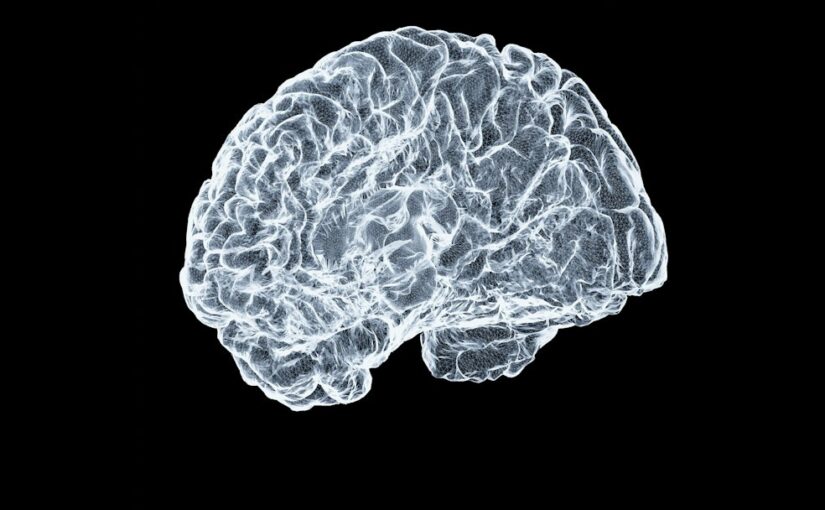
Neuroimaging studies repeatedly identify differences in several regions in people with ADHD compared with control groups. The most robust structural findings involve the prefrontal cortex, basal ganglia (including the caudate and putamen), cerebellum, and parts of the limbic system. These differences are usually seen as smaller volumes or delayed maturation rather than gross structural abnormalities.
| Domain | Typical structural differences | Clinical relevance |
|---|---|---|
| Attention and executive control | Reduced volume or delayed cortical thinning in prefrontal regions | Linked to inattentive symptoms and planning difficulties |
| Motor and inhibition | Smaller basal ganglia structures (caudate, putamen) | Associated with impulsivity and motor restlessness |
| Timing and coordination | Smaller cerebellar volumes or altered cerebellar cortex | May affect timing, coordination, and some cognitive skills |
| Emotion regulation | Structural variations in limbic areas (amygdala, hippocampus) | Related to emotional lability and mood comorbidity |
| Connectivity implications | Altered white matter tracts connecting frontal and subcortical regions | Explains inefficient network communication and processing speed |
How do these structural differences relate to ADHD symptoms?
Structural differences provide a biological context for the cognitive and behavioral profile of ADHD. Prefrontal cortical differences support difficulties with attention control, working memory, planning, and impulse control. Basal ganglia variations are linked to response selection and motor regulation, while cerebellar differences may contribute to timing and coordination problems.
Importantly, anatomical differences are not deterministic. Many people with structural variations function well with appropriate supports and treatment. Structural findings offer one piece of the puzzle that complements behavioral assessment and functional measures such as task-based or resting-state functional MRI.
How do brain differences change across the lifespan in ADHD?
Brain structure differences in ADHD often reflect altered developmental trajectories rather than fixed deficits. Longitudinal studies show delayed cortical maturation in some regions, most notably in prefrontal areas responsible for executive control. That means cortical thinning or volume reductions observed in childhood may normalize or converge gradually with age for some individuals.
Some subcortical differences, like smaller basal ganglia volumes, may persist into adolescence or adulthood in many people. At the same time, the clinical expression of ADHD changes with age: hyperactivity often decreases, while inattentive and executive challenges can remain. Structural imaging helps explain why symptoms shift over time but does not reliably predict individual outcomes on its own.
What factors influence brain development in ADHD?
Multiple factors interact with genetic predisposition and brain development. Prenatal exposures, early life stress, sleep disturbances, nutrition, and comorbid conditions like anxiety or learning disorders all influence brain maturation. Medication exposure such as stimulant treatment has been studied for potential effects on brain structure; current evidence indicates some normalization of differences in treated samples, but findings vary and more research is needed.
Can brain imaging diagnose ADHD or guide personalized treatment?
At present, structural brain imaging cannot diagnose ADHD reliably at the individual level. Group-level differences are robust in research but show too much overlap between people with and without ADHD to serve as diagnostic biomarkers. Clinical diagnosis remains behavioral, based on history, rating scales, and functional impairment.
However, imaging can inform research on mechanisms and suggest targets for interventions. For example, studies linking fronto-striatal circuitry to impulsivity support treatments that strengthen executive skills, either through medication, cognitive training, or behavioral programs. Functional imaging and connectivity analyses sometimes indicate patterns that correlate with response to a particular treatment, but these techniques are not yet routine in clinical practice.
What do structural differences imply for treatment strategies?
Understanding structure-function relationships helps prioritize interventions. When prefrontal differences align with executive dysfunction, interventions that scaffold organization, break tasks into smaller steps, and target working memory can be effective. If basal ganglia and motor-related differences are dominant, behavioral strategies that reduce impulsive responding and increase physical outlets for activity may help.
Pharmacological treatments, especially stimulant medications, have a strong evidence base for reducing core ADHD symptoms. Nonpharmacological approaches including parent training, classroom accommodations, cognitive behavioral therapy, and structured routines are essential complements. Tailoring strategies to the individual symptom profile and functional needs yields the best outcomes.
How do structural findings connect with brain connectivity patterns?
Structural differences often co-occur with altered connectivity between brain regions. White matter tracts that connect prefrontal cortex to subcortical and parietal regions can show differences in integrity, which relates to slower information transfer and less efficient network coordination. For more on connectivity evidence, see research on brain connectivity patterns in ADHD that complements structural findings.
Which cognitive tests and clinical assessments reflect structural effects?
Neuropsychological tests for attention, inhibition, processing speed, and working memory correlate with structural findings in many studies. For example, tests that measure sustained attention and response inhibition often show associations with prefrontal and basal ganglia measures. Standardized rating scales and functional impact reports remain essential because they capture real-world impairment that imaging alone cannot determine.
Examples of assessments
Common tools include continuous performance tests, digit span and other working memory measures, and standardized behavior rating scales for home and school. Combining these assessments with developmental and medical history provides a reliable clinical picture without needing imaging for most patients.
How should clinicians, parents, and adults interpret imaging results?
If structural imaging is available, interpret results cautiously. Radiological findings such as minor volume differences or nonspecific variations do not confirm or rule out ADHD. Imaging is most useful to exclude alternative neurological conditions when clinical history or examination raises concerns, for example when there is a new focal neurological deficit, seizures, or atypical developmental regression.
Discuss imaging results with a neurologist, psychiatrist, or neuropsychologist who understands both structural findings and their limits. Treatment decisions should remain grounded in behavioral and functional assessments, not imaging alone.
What practical strategies follow from understanding brain structure differences?
Knowing which brain systems are implicated helps shape targeted supports. For attention and executive issues, use external structure: visual schedules, timers, checklists, and clear step-by-step instructions. For hyperactivity and impulsivity, add structured movement breaks, clear behavioral expectations, and consistent reinforcement.
Adults can adopt strategies such as setting up environmental cues, using digital task management tools, and planning for when executive resources are low. For children, combining parent training and school accommodations delivers measurable gains while supporting brain development.
How can habit formation and routine support brain function in ADHD?
Consistent routines reduce cognitive load and leverage habit memory circuits that can bypass weaker executive control during busy moments. Habit formation strategies are practical complements to interventions aimed at executive deficits. For concrete habit-building techniques, see guidance on habit formation strategies for ADHD, which provide step-by-step methods to build productive routines.
What special considerations apply to children with ADHD?
Children show the most pronounced maturational differences in cortical development, so early interventions that support skill acquisition and adaptive routines are especially valuable. Fostering strengths, interests, and motivation helps children engage in tasks that build executive capacity over time. Resources that focus on strengths can improve self-esteem and long-term outcomes. For practical approaches to nurture abilities, consider material on encouraging strengths and interests in ADHD children.
Educational and parental strategies
Classroom accommodations such as seat placement, chunked assignments, and frequent feedback reduce demands on developing systems. Parent coaching to implement consistent routines, clear consequences, and structured rewards supports brain maturation and daily functioning.
What does the research say about medication and structural brain changes?
Studies that examine medication effects on brain structure report mixed findings. Some longitudinal research suggests that stimulant treatment may be associated with partial normalization of certain brain differences, particularly in fronto-striatal circuits, while other studies show minimal or inconsistent effects. Medication remains evidence based for symptom reduction, but imaging alone should not be used to judge treatment success.
Clinical monitoring should focus on symptom change, side effects, functioning in daily life, and developmental milestones rather than imaging endpoints.
Are there credible biomarkers for ADHD yet?
Researchers are actively searching for biomarkers that could support diagnosis, prognosis, or treatment selection. So far, no single structural measure functions as a biomarker with sufficient sensitivity and specificity for clinical use. Multimodal approaches that combine imaging, cognitive testing, genetics, and behavioral data show promise in research settings but are not yet standard practice.
How might future research change practice?
Advances in machine learning applied to large, well-characterized datasets may yield predictive models that can inform individualized care. Progress depends on rigorous replication, diverse samples, and careful evaluation of clinical utility. Until then, imaging remains primarily a research tool for understanding mechanisms and heterogeneity within ADHD.
Examples and expert-backed context
Key large-scale studies illustrate current knowledge. A major mega-analysis pooled structural MRI data from thousands of participants and reported smaller volumes in several subcortical regions in people with ADHD. Longitudinal work by developmental neuroscientists reported a delay in cortical maturation in ADHD compared with peers, particularly in regions supporting executive function. These high-quality studies support the interpretation that ADHD involves altered developmental timing and regional volumetric differences rather than a single uniform brain lesion.
For consolidated information from a trusted source on how brain research informs ADHD understanding, see the NIMH overview of ADHD and brain research. This resource summarizes current findings and their clinical implications.
How should families and adults act on this information?
Practical steps include obtaining a thorough clinical assessment if ADHD is suspected, focusing on behavioral and functional evidence when making treatment decisions, and using targeted interventions to address executive deficits and behavioral challenges. Consider combined approaches: evidence-based medication when appropriate, structured behavioral supports, and skills training for organization and time management.
Engage professionals for diagnostic clarity when comorbid conditions are present or when symptoms are severe. Use strengths-based strategies to support motivation and resilience, and maintain regular follow-up to adapt interventions as needs change.
FAQ
Can a brain scan confirm if someone has ADHD?
No. Brain scans show group-level differences but cannot confirm ADHD in an individual because findings overlap substantially with typical development.
Do structural brain differences mean ADHD is a disease?
Structural differences indicate neurodevelopmental variation. They are not a disease label, but they reflect biological contributions to behavior and cognition. ADHD is a neurodevelopmental condition defined by behavior and functional impairment.
Will medication change brain structure in ADHD?
Some studies suggest medication may influence brain development modestly, but evidence is mixed. Treatment decisions should be based on symptoms, functioning, and side effect profiles.
Should every child with ADHD get an MRI?
No. Routine MRI is not recommended for typical ADHD. Imaging is reserved for atypical presentations or when another neurological concern is suspected.
Practical next steps
If you suspect ADHD, start with a comprehensive clinical evaluation that includes developmental history, behavior rating scales, and functional assessment. Use findings about brain structure differences to inform supportive interventions that target specific cognitive and behavioral challenges. For parents and educators, implement structured routines, explicit instructions, and environmental supports that scaffold executive demands. For clinicians, stay informed about emerging multimodal research while relying on behavioral criteria and functional outcomes to guide care.
Bibliography
- Hoogman M, Bralten J, Hibar DP, et al. Subcortical brain volume differences in participants with attention deficit hyperactivity disorder in children and adults: a cross-sectional mega-analysis. Lancet Psychiatry. 2017;4(4):310-319. PubMed. https://pubmed.ncbi.nlm.nih.gov/28248326/
- Shaw P, Eckstrand K, Sharp W, et al. Attention-deficit/hyperactivity disorder is characterized by a delay in cortical maturation. Proceedings of the National Academy of Sciences. 2007;104(49):19649-19654. PubMed. https://pubmed.ncbi.nlm.nih.gov/17460041/
- National Institute of Mental Health. Attention-Deficit/Hyperactivity Disorder (ADHD). NIMH. https://www.nimh.nih.gov/health/topics/attention-deficit-hyperactivity-disorder-adhd
- Centers for Disease Control and Prevention. Data and statistics about ADHD. CDC. https://www.cdc.gov/ncbddd/adhd/
- American Psychiatric Association. Diagnostic and Statistical Manual of Mental Disorders, Fifth Edition. Arlington, VA: American Psychiatric Publishing; 2013.